[O-2-173] Stripe sign of precentral gyri in amyotrophic lateral sclerosis: a novel finding on PADRE
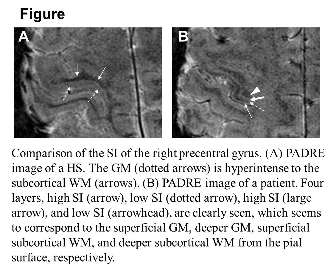
Purpose: To evaluate the signal-intensity (SI) of the precentral gyri (PG) in the amyotrophic lateral sclerosis (ALS) patients in comparison with healthy subjects (HS) by using a phase-weighted MR imaging technique “Phase Difference Enhanced Imaging (PADRE)”. Methods: The PADRE was performed in 5 ALS patients and 20 HS at 3T MRI system. For the PG on PADRE images, two radiologists compared the SI of the grey matter (GM) with the subcortical white matter (WM) and divided into 3 grades: hyperintense, isointense, or hypointense. Results: The GM of the PG was hyperintense to the subcortical WM in all 20 HS, and hypointense in all 5 ALS patients (Fig.). Furthermore, in ALS patients, four layers, high, low, high, and low SI from the pial surface of the PG, were seen on PADRE images, which seemed to correspond to the superficial GM, deeper GM, superficial subcortical WM, and deeper subcortical WM, respectively (stripe sign) (Fig. B). Conclusions: In ALS patients, the PADRE images showed the four layers in the PG, which might reflect the iron deposition in a part of the GM and the reduced concentration of myelin in the superficial subcortical WM.