[O-2-250] Situation of arteriolar elasticity obtained from spin-echo signal fluctuations in the human brain
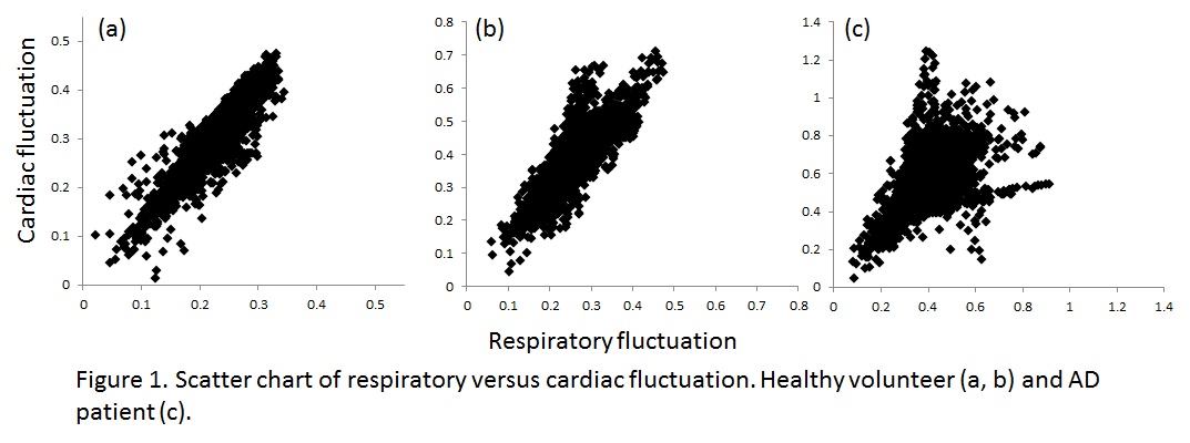
[Introduction] Arteriolar elasticity (AE) may predict the progress of dementia. Focusing on arteriolar vasomotion appearing in MR signal fluctuation, we propose a new method to obtain AE deterioration. [Materials & Methods] SE-EPI was performed under 1.5-T MRI for 2 healthy volunteers and an AD patient. The time series data were Fourier-transformed to map the spectral intensities in respiratory (R:0.2-0.5 Hz) and cardiac (C:0.8-1.2 Hz) ranges. The scatter charts of R and C values in pixels were made. [Results] The scatter charts of volunteers (Fig. 1a,b) show good correlation (r=0.89 & 0.88) while that of AD patient (Fig. 1c) scatters (r=0.52). [Discussion]The fluctuation in R range reflects regional vasomotion caused by PaCO2 changes while that in C range reflects regional vasomotion and global blood flow changes caused by periodic blood pressure. When AE deteriorates regionally, correlation of regional vasomotion and global blood flow decreases: this appears a decrease in correlation of the scatter chart. Our results show good correlation for volunteers and poor correlation for AD patient. [Conclusion] Situation of AE can be obtained by analyzing SE signal fluctuation.