[O-3-276] The discrepancy between pseudocontinuous ASL and CE-MRI of primary intra-axial brain tumor
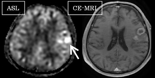
INTRODUCTION: This study was performed to investigate the discrepancy between pseudocontinuous arterial spin labeling (pCASL) MRI and contrast-enhanced (CE) MRI findings of primary intra-axial brain tumor. MATERIALS AND METHODS: Thirty consecutive cases with intra-axial brain tumor (6 metastasis, 5 low grade glioma (LGG), 13 high grade glioma (HGG), 6 primary CNS lymphoma (PCNSL)) underwent pCASL and CE-MRI were enrolled. Imaging findings were divided into 3 groups; 1) match: hyperintensity on pCASL was included in the enhanced area on CE-T1WI, 2) mismatch; pCASL-hotspot was found outside of the enhanced area, 3) controversial: difficult to classified into 1) or 2).RESULTS: Six cases were eliminated because of poor image quality. Fifteen, seven and two cases were classified into match, mismatch, and controversial, respectively. The histology of mismatch cases were 4: LGG, 2: HGG, 1: PCNSL. The controversial cases were HGG and PCNSL cases. All pCASL images of metastasis cases were classified into match group.CONCLUSIONS: “ASL CE-MRI mismatch” indicate glioma or PCNSL. This finding is useful in diagnosis of primary intra-axial brain tumor.